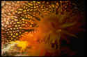
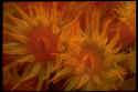
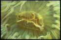
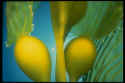
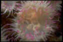
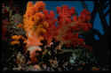
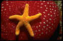
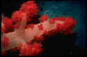
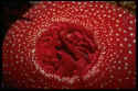
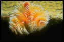
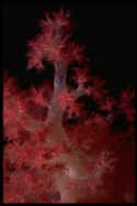
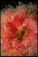
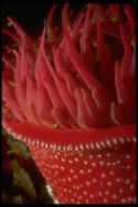
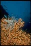
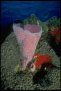
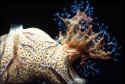
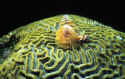
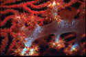
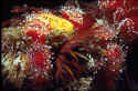
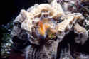
underwater photography of great white sharks, dolphins, divers and other marine life by Stephen Brunson
underwater photography of great white sharks, dolphins, divers and other marine life by Stephen Brunson underwater photography of great white sharks, dolphins, seals, divers and other marine creatures underwater photography of great white sharks and dolphins by Stephen Brunson Award winning underwater photography of great white sharks and dolphins by Stephen Brunson All about underwater photographer Stephen Brunson underwater photography of great white sharks, dolphins, divers and other marine life by Stephen Brunson free screensavers, free wallpaper, free animations, free images Underwater links and reciprocal link exchange
Anemones and Corals
Limited Edition Signed color photography for sale by Stephen Brunson.
Stock photography for publication, commercial use and all other media for sale.

Click on picture for more detail

The anemones body cavity is divided into sacs, septa, extending from the body wall      The anenomes septa increase the surface available for the secretion of digestive juices and the absorption of nourishment. They also contain the gonads which contain the sperm and eggs      The anenomes eggs are usually fertilized in the gastric cavity, and the young are discharged from the mouth as free-swimming larvae, which soon attach themselves to surfaces      I photographed this kelp plant in La Jolla Cove at 1:2 macro with a sunburst in the background at F22, with dual opposing strobes. Kelp is the common name for large, leafy brown algae known as seaweed, of the order Laminariales, that grow along colder coastlines      Hermit crabs sometimes allow anenomes to attach themselves to their shells, for protection

I photographed this picture in Fiji, the soft coral capital of the world. In this photograph, there in not only soft coral, but precious black coral, elkhorn coral, and a giant clam. Coral is the common name for a large group of marine invertebrates of the class Anthozoa, which also includes sea anemones, in the phylum Coelenterata      This picture was taken in the Chanel Islands off California, where small and large anemones flourish in many different colors      I photographed this picture in Fiji, the soft coral capital of the world. I took this one with a 1:3 macro extension tube, in the evening, managing to get a partial sunburst at F22.      Notice the sea urchin stabbing the anemone with one of his spines. This picture was taken in the Chanel Islands off California, where small and large anemones flourish in many different colors      I photographed this picture in Bonaire, with a 1:1 macro extension tube. Bonaire is the macro capital of the world, with many varieties of christmas tree coral. True corals secrete calcium carbonate from the bottom half of the stalk of the individual animal, or polyp, forming skeletal cups to which the polyps are anchored and into which they withdraw for protection

I photographed this picture in Fiji, the soft coral capital of the world. I took this one with a 1:1 macro extension tube, at night, with opposing strobes. At night the tentacles extend from the cup, seize animal plankton that wash against them, and carry the food to the mouth. Stinging cells, or nematocysts, on the tentacles can also paralyze prey      The strawberry anenome, Corynactis australis, is usually found in cooler temperate oceans, this one in the northern Pacific Ocean      The sea anenomes body is cylindrical: one end is closed, which it attaches to rocks, coral, and even crustaceans, with a slitlike mouth and tentacles on the other end      I photographed this picture in Palau, the seventh underwater wonder of the world. It is a giant orange seafan, with a black and white Crynoid on it, quite common in the south Pacific      I photographed this picture in Cozumel, part of the second largest great barrier reef in the world. This is an Azure Vase Coral, which is very common in the Caribbean, and Cozumel

Some scleractinians exist as solitary polyps, such as Fungia of the Indian and Pacific oceans, but the majority are colonial. Most colonial polyps average from 1 to 3mm in diameter. They are connected laterally by tubes that are simply an extension of the polyps' gastrovascular cavities, and the colony grows by asexual budding from the base or the oral disc of the polyps      I photographed this picture in Roatan, which is part of the second largest great barrier reef in the world. This is a christmas tree coral on a massive brain coral, shot at approximately 1:2 macro.      I photographed this picture in Fiji at night, at 1:1 macro, of soft coral with a red giant gorgonian in the background.      Notice the lemon yellow nudibranch next to the strawberry anemones. This picture was taken in the Chanel Islands off California, where small and large anemones flourish in many different colors      This one I photographed in Cozumel at night, at 1:2 macro. What I noticed about this image was the contrast between the soft tentacles, or featherduster, with the hard calcium carbonate coral in the background.

BrunsonImages@att.net